MSK 2023 Joint Meeting With Interactive Course on axSpA
From October 8th to 13th, the MSK 2023 Global Musculoskeletal Societies Joint Meeting took place in London under the auspices of the International Skeletal Society (ISS). The MSK Refresher Course was an anchor of the program as usual. As a part of it, on October 12th, an interactive course on axial spondyloarthritis (axSpA) took the stage as our very own highlight. It’s time for a recap.
Specialty Societies from Around the World at the MSK 2023 Joint Meeting
With over 1,000 experts from radiology, orthopedics, and pathology from around the world coming together, the MSK 2023 Global Musculoskeletal Societies Joint Meeting offered a unique platform for the exchange of knowledge and discussion of cases in the field of musculoskeletal disorders. Every year, the event takes place in a different location. This time, the international congress made a stop in London.

The International Skeletal Society (ISS) invites musculoskeletal (MSK) societies to London. — photo: Unsplash — Benjamin Davies
For the 50th anniversary of the International Skeletal Society, the event featured an impressive array of presentations and discussions uniting #allMSKsocieties under one hashtag. The BerlinCaseViewer was also part of it, serving as the technical backbone.
Enhancing the Experience with the BCV Web App
Participating in the MSK 2023 Joint Meeting was truly special for us. After all, this globally esteemed event brings together the brightest minds in the field of musculoskeletal disorders. The fact that the International Skeletal Society celebrated its 50th anniversary this year added even more significance to the event. And it was up to us — or rather, our web app — to pave the way for in-depth interdisciplinary discussions.
And here’s how it all started. A course on axial spondyloarthritis, organized by radiologists Robert G. Lambert and Kay G. Hermann, leveraged the innovative BerlinCaseViewer platform to showcase cases. The plan included contributions from a total of 9 presenters from around the world, featuring intriguing and illustrative case examples. Everything was meticulously orchestrated down to the last minute.

9 presentations on axial spondyloarthritis were on the agenda.
Participants, on the other hand, were encouraged to actively engage by studying the findings in detail and answering multiple-choice questions. This way, we could convey specialized knowledge as well as provide an overview-style introduction to the subject of axSpA.
What You Might Have Missed at this MSK Refresher Course
Kay G. Hermann from our team kicked off the course with a technical introduction to the web version of BerlinCaseViewer. Doing so, he had a giraffe from Kenya in tow. Watch it again here:
This is how the BerlinCaseViewer web app works — using a giraffe as an example. Yes, you read that right. Giraffe.
Following the motto “Bringing axSpA into focus,” the course continued with Kay G. Hermann sharing radiological insights about axial spondyloarthritis and its various manifestations at the sacroiliac joints. Brazilian radiologist Clarissa Canella then picked up this thread to discuss typical radiological signs of axSpA and how to utilize them for diagnosis and classification.
You might remember, in 2009, there were several changes in the classification of inflammatory spinal diseases that led to a new nomenclature. More on this in the interview with rheumatologist Joachim Sieper.
Important Differential Diagnoses at a Glance
Next up, Dutch radiologist Monique Reijnierse delved into key differential diagnoses to explore how axSpA distinguishes itself from other musculoskeletal conditions such as osteoarthritis, osteitis condensans, fractures, and bacterial inflammation.
In a similar vein, but paying closer attention to young patients, Canadian radiologist Jacob Jaremko pondered questions like, “How do children differ from adults? What growth-related MRI phenomena should be considered?”
Differential diagnoses, especially inflammatory patterns visible in imaging, were of primary interest to radiologist Winston Rennie from Loughborough University near Leicester, England.

The BerlinCaseViewer highlights essential differential diagnoses.
Due to the ongoing Israel-Gaza conflict, Iris Eshed from Sheba Medical Center in Tel Aviv had to cancel her participation at short notice. In her stead, Kay G. Hermann summarized her presentation on additional differential diagnoses for the attendees, covering osteomyelitis, the SAPHO syndrome, and diffuse idiopathic skeletal hyperostosis, or DISH.
Reading the Signs: Is it Truly axSpA? What Alters with Treatment?
Radiologist Robert Lambert from Canada tackled complex case examples in his presentation, addressing a crucial question: How can we establish a clear diagnosis in situations where both syndesmophytes, indicating axSpA, and degenerative processes are present?
Danish rheumatologist Mikkel Østergaard also utilized exemplary cases from his clinical practice to investigate the role of MRI in follow-up assessments. He explored how often imaging should be used to identify treatment failures, and how the patterns of findings can evolve and change over time.

Providing accurate answers, making the right diagnosis.

Faculty of the course (in part): Diekhoff, Reijnierse, Lambert, Canella, Hermann, Østergaard.
Technological Innovations to Broaden Radiological Horizons
Radiologist Torsten Diekhoff from Charité — Universitätsmedizin Berlin took us on a deeper dive into the technical realm. He unveiled cutting-edge imaging approaches and their pivotal role in diagnosis. With real-life examples, he showcased the novel possibilities in modern radiology, such as simulating a computed tomography image using an MRI machine, completely eliminating the need for X-ray radiation. Conversely, you can gauge the level of inflammation at the sacroiliac joints with a CT scan, especially when an MRI isn’t at your fingertips or when a CT scan is necessary for other reasons.
Over 250 participants eagerly pored over the presented cases, all with the assistance of the BerlinCaseViewer web app. For technical support, Friedrich Hermann, Kay G. Hermann’s son, joined forces with the BerlinCaseViewer team.
Now it’s your turn! Examine the case that Kay Hermann showed in the course. There are three embedded questions. After answering each question, you will receive further information about the case.
Our heart beats for imaging in rheumatology. We regularly run online and on-site courses. When will you be attending one of our courses?
Download BerlinCaseViewer
Related posts:
 https://berlincaseviewer.de/wp-content/uploads/Chest-X-ray-ENG-02.jpg
1080
1920
Eva Reidemeister
https://berlincaseviewer.de/wp-content/uploads/2020/05/bcv-logo-1.svg
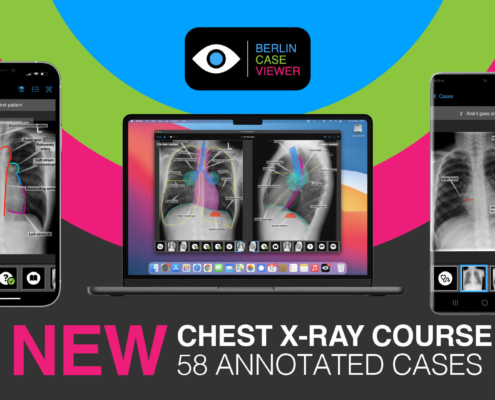
Eva Reidemeister2023-11-07 19:42:232023-11-08 13:44:01#IDoR2023: Brandnew Chest X-ray Module Celebrating International Day of Radiology
https://berlincaseviewer.de/wp-content/uploads/Chest-X-ray-ENG-02.jpg
1080
1920
Eva Reidemeister
https://berlincaseviewer.de/wp-content/uploads/2020/05/bcv-logo-1.svg
Eva Reidemeister2023-11-07 19:42:232023-11-08 13:44:01#IDoR2023: Brandnew Chest X-ray Module Celebrating International Day of Radiology https://berlincaseviewer.de/wp-content/uploads/MRI-superbreit3.jpg
500
1500
Eva Reidemeister
https://berlincaseviewer.de/wp-content/uploads/2020/05/bcv-logo-1.svg
Eva Reidemeister2023-08-04 13:45:102023-08-04 13:55:13NEW: Module on MRI of the Knee Joint Focussing on Meniscus Injuries
https://berlincaseviewer.de/wp-content/uploads/MRI-superbreit3.jpg
500
1500
Eva Reidemeister
https://berlincaseviewer.de/wp-content/uploads/2020/05/bcv-logo-1.svg
Eva Reidemeister2023-08-04 13:45:102023-08-04 13:55:13NEW: Module on MRI of the Knee Joint Focussing on Meniscus Injuries https://berlincaseviewer.de/wp-content/uploads/Polish-combo.png
1200
1920
Kay G. Hermann
https://berlincaseviewer.de/wp-content/uploads/2020/05/bcv-logo-1.svg
Kay G. Hermann2023-07-12 09:53:342023-07-20 08:39:31Arthritis in Polish: Our New Learning Module
https://berlincaseviewer.de/wp-content/uploads/Polish-combo.png
1200
1920
Kay G. Hermann
https://berlincaseviewer.de/wp-content/uploads/2020/05/bcv-logo-1.svg
Kay G. Hermann2023-07-12 09:53:342023-07-20 08:39:31Arthritis in Polish: Our New Learning ModuleThis post is also available in: German

