NEW: Module on MRI of the Knee Joint Focussing on Meniscus Injuries
The diagnosis of knee injuries raises several questions: Is the injury the result of an accident or the result of degenerative changes in the knee joint? At this point, after anamnesis and clinical examination, medical imaging comes into play.
Magnetic resonance imaging (MRI) can be used to get a clear picture of the injury. Our new module on MRI of the knee joint focussing on meniscus injuries helps you to interpret the radiological images correctly to avoid misinterpretations.

Dr. Wolfgang Fischer
Meniscus injuries from bucket-handle tear to flap tear to root tear
You are already familiar with the anatomy of the knee joint. Now it’s time to explore different patterns of meniscus injuries. These include longitudinal, transverse, and horizontal tears, as well as various combinations of these types. Our new radiology module showcases the key meniscus injuries through eight pathological cases and two companion cases:
- Bucket-handle tear: It is the result of a long longitudinal tear. When the torn fragment shifts into the notch, the configuration of a “bucket handle” is created.
- Flap tear: Flap tears are usually caused by a combination of longitudinal and transverse tears. The detached piece (“flap”) can then turn over and cause entrapment. These flaps are often found at or below the base of the meniscus and adjacent to the tibial plateau or femoral condyle.
- Root tear: A root tear is directly at or near the bony anchorage of the meniscus to the tibial plateau (“meniscus root”).
In addition, there are various meniscus changes that can be attributed to degenerative processes in the knee. With our module, you get an overview and can sharpen your eye for the subtle differences in imaging.

Collaboration with Wolfgang Fischer
The new module was created in collaboration with the radiologist Wolfgang Fischer, who specializes in musculoskeletal radiology. What Wolfgang Fischer shares with the BerlinCaseViewer is above all his enthusiasm for interactive teaching and learning. Maybe you know his own app, “MRI-Essentials”.
His app is based on the textbook “MRI-Essentials.com”, which is known worldwide as a relevant work on MRI of the musculoskeletal system, specializing in orthopedics and sports medicine. This is not least due to international co-authors such as Rick Kijowski and Ali Guermazi, who are themselves experts in musculoskeletal radiology respectively osteoarthritis. “MRI-Essentials”, the app, is available in English and German. With an annual subscription of just under 40 Euro, you can unlock all the images it contains.
New module for the BerlinCaseViewer
Wolfgang Fischer is an active member of radiological societies like the ESSR and ISS. At congresses, he regularly shares his knowledge. As a dedicated speaker, he puts a great deal of effort and attention to detail into the preparation of his presentation slides, some of which are illustrated with graphics he has drawn himself. Ultimately, that is what brought us together, because his anatomical drawings and our colored overlays complement each other perfectly.

The radiologists Kay G. Hermann and Wolfgang Fischer 2022 in Barcelona
Wolfgang Fischer has prepared eight selected cases for the BerlinCaseViewer. The resulting module is intended as an in-depth unit for everyone who is already familiar with “MRI-Essentials”, but also offers a good introduction to orthopedic musculoskeletal radiology.
Find the “MRI-Essential Series” in the BCV app!
For 7.99 Euro you get an exciting overview module on meniscus injuries, including high-resolution MRI images with colored annotations, multiple-choice questions and tips for diagnosis. Some cases also contain surgical photos, surgical videos or follow-up examinations.
You just want to see what the app has in store before you download for more? No problem. We have selected one of two bonus cases for you to try out in your web browser: A 24-year-old man who suffered a medial collateral ligament injury two years ago has been experiencing pain while playing soccer for about two weeks.
Discover the MRI changes, the surgical video and tips for learning here in the interactive web version:
You can find more about this and other exciting cases in our app.
About the author
After studying medicine at the Universities of Heidelberg (Germany), Geneva (Switzerland) and Montpellier (France), Wolfgang Fischer worked from 1992 to 2008 in the Department of Diagnostic Radiology and Neuroradiology at the Augsburg municipal hospital in Germany. In 1999, he was a visiting scholar at the University of California in San Francisco (USA). Since 2008 he has been working in a highly specialized MRI department in an orthopedic hospital. In 2018, he was awarded the Eugenie-und-Felix-Wachsmann prize by the German radiological society for merits in radiologic education.
Download BerlinCaseViewer
Related posts:
 https://berlincaseviewer.de/wp-content/uploads/Rheuma-Day-2024-Header-WP.jpg
843
1500
Eva Reidemeister
https://berlincaseviewer.de/wp-content/uploads/2020/05/bcv-logo-1.svg
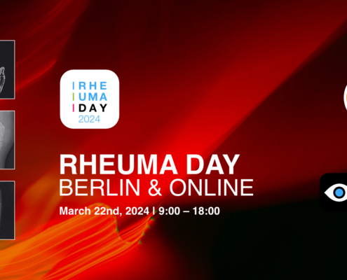
Eva Reidemeister2024-02-13 16:36:512024-02-13 16:36:51Clinical Insights: Rheuma Day 2024 — Focussing on MSK Radiology
https://berlincaseviewer.de/wp-content/uploads/Rheuma-Day-2024-Header-WP.jpg
843
1500
Eva Reidemeister
https://berlincaseviewer.de/wp-content/uploads/2020/05/bcv-logo-1.svg
Eva Reidemeister2024-02-13 16:36:512024-02-13 16:36:51Clinical Insights: Rheuma Day 2024 — Focussing on MSK Radiology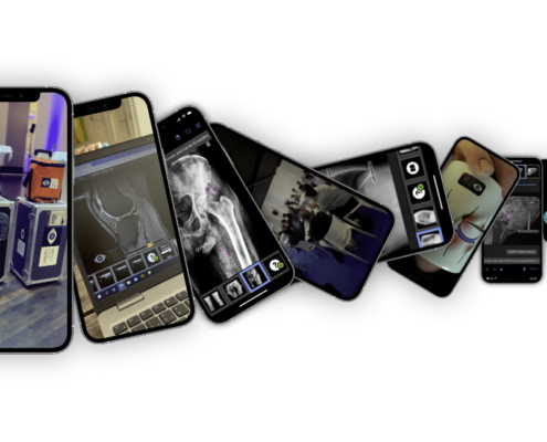 https://berlincaseviewer.de/wp-content/uploads/Year-in-Review-2023.png
650
1300
Eva Reidemeister
https://berlincaseviewer.de/wp-content/uploads/2020/05/bcv-logo-1.svg
Eva Reidemeister2023-12-31 09:47:552024-01-01 16:41:49Rays of Progress: A Radiology Learning Journey Through 2023
https://berlincaseviewer.de/wp-content/uploads/Year-in-Review-2023.png
650
1300
Eva Reidemeister
https://berlincaseviewer.de/wp-content/uploads/2020/05/bcv-logo-1.svg
Eva Reidemeister2023-12-31 09:47:552024-01-01 16:41:49Rays of Progress: A Radiology Learning Journey Through 2023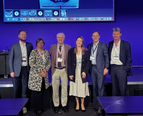 https://berlincaseviewer.de/wp-content/uploads/ISS-2023-faculty-landscape-scaled.jpeg
1616
2560
Eva Reidemeister
https://berlincaseviewer.de/wp-content/uploads/2020/05/bcv-logo-1.svg
Eva Reidemeister2023-11-03 22:06:362023-11-04 01:54:13MSK 2023 Joint Meeting With Interactive Course on axSpA
https://berlincaseviewer.de/wp-content/uploads/ISS-2023-faculty-landscape-scaled.jpeg
1616
2560
Eva Reidemeister
https://berlincaseviewer.de/wp-content/uploads/2020/05/bcv-logo-1.svg
Eva Reidemeister2023-11-03 22:06:362023-11-04 01:54:13MSK 2023 Joint Meeting With Interactive Course on axSpAThis post is also available in: German

