#IDoR2023: Brandnew Chest X-ray Module Celebrating International Day of Radiology
On November 8th, 1895, the future Nobel laureate Wilhelm Conrad Roentgen had a stroke of luck: Radiation invisible to the human eye had the ability to make otherwise concealed things visible. Initially, Roentgen referred to these short-wavelength, high-energy rays as “X-rays.” Later on, in German they would be called “Röntgenstrahlen” (Roentgen rays) in his honor. Among the first images was an X-ray of his wife’s hand, with a ring seemingly floating around her bony finger.
For the 11th International Day of Radiology on November 8th, we celebrate not only the discovery of X-rays, but also the impressive progress made in radiology since then.
Fascinating Chest X-rays
Thanks to X-rays, we can now not only visualize finger bones beneath tissues but also the lungs, the entire mediastinum including ribs, heart, and spine within the body. Additionally, X-rays can reveal materials introduced into the body, such as central catheters, pacemakers, or ports.
The imaging of the heart and lungs through X-rays is quintessential in radiology. It is one of the oldest fields of medical imaging, with numerous accomplishments. Just think about the containment of tuberculosis (TB) in past decades!

A mobile X-ray unit during healthcare support for the rural population in Hermstedt in the GDR (German Democratic Republic) — photo: Federal Archive
From the Early Detection of Tuberculosis Onwards
In some countries, mobile X-ray units were even built into buses or trains to combat the TB epidemic. Chest X-rays provided crucial diagnostic clues. In Germany, the home of BerlinCaseViewer, tuberculosis has become relatively rare, and large-scale X-ray screenings are a thing of the past. However, globally, tuberculosis remains one of the top ten leading causes of death.1 And X-ray technology continues to be relevant to this day.
Check out the case preview in the web version.
Thoracic Radiology Made Easy — New Module
Whether in general practice, anesthesia, internal medicine, or intensive care — in this part of the world, chest X-rays remain an essential routine diagnostic tool, especially for assessing pneumonia or pulmonary edema.
There is a plethora of learning materials for interpreting chest X-rays. With our new module “Thoracic Imaging: Chest X-ray Essentials”, we are by no means the first. However, we aim to be the best and perhaps the most amusing in the field. With a wealth of expertise and a touch of humor, we take you into the fascinating world of heart and lung X-ray images to acquaint you with thoracic radiology.

The latest module from BerlinCaseViewer offers a total of 58 cases, meticulously annotated. Concise learning texts and engaging multiple-choice questions guide you through the selected cases, highlighting the key aspects.
In collaboration with Torsten Diekhoff from the Department of Radiology at Charité — Universitätsmedizin Berlin and the dedicated students Niclas Doepner, Patrick Hoffacker, and Mohammad Asif Iqbal, we have created a three-part course designed to convey essential knowledge not only to medical students but also to all other interested individuals.
Compact. Easy. Interactive. In one app. With the BerlinCaseViewer, you have “Thoracic Imaging: Chest X-ray Essentials” at your fingertips, even when there is no internet connection available.
In Time for the 11th International Day of Radiology
This learning experience is available in German (🇩🇪) and English (🇬🇧) in our app starting from November 8th.
Discover “Thoracic Imaging: Chest X-ray Essentials”
In the BerlinCaseViewer App Now
Related posts:
 https://berlincaseviewer.de/wp-content/uploads/Rheuma-Day-2024-Header-WP.jpg
843
1500
Eva Reidemeister
https://berlincaseviewer.de/wp-content/uploads/2020/05/bcv-logo-1.svg
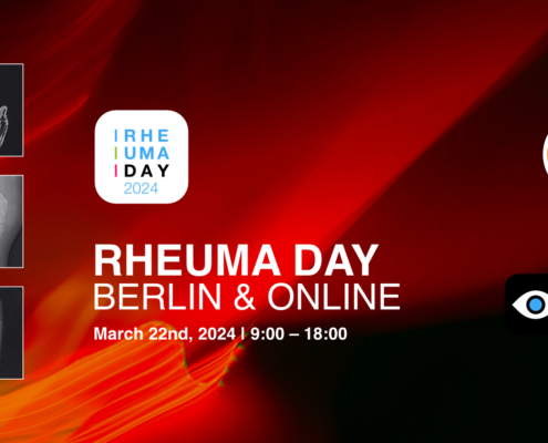
Eva Reidemeister2024-02-13 16:36:512024-02-13 16:36:51Clinical Insights: Rheuma Day 2024 — Focussing on MSK Radiology
https://berlincaseviewer.de/wp-content/uploads/Rheuma-Day-2024-Header-WP.jpg
843
1500
Eva Reidemeister
https://berlincaseviewer.de/wp-content/uploads/2020/05/bcv-logo-1.svg
Eva Reidemeister2024-02-13 16:36:512024-02-13 16:36:51Clinical Insights: Rheuma Day 2024 — Focussing on MSK Radiology https://berlincaseviewer.de/wp-content/uploads/Year-in-Review-2023.png
650
1300
Eva Reidemeister
https://berlincaseviewer.de/wp-content/uploads/2020/05/bcv-logo-1.svg
Eva Reidemeister2023-12-31 09:47:552024-01-01 16:41:49Rays of Progress: A Radiology Learning Journey Through 2023
https://berlincaseviewer.de/wp-content/uploads/Year-in-Review-2023.png
650
1300
Eva Reidemeister
https://berlincaseviewer.de/wp-content/uploads/2020/05/bcv-logo-1.svg
Eva Reidemeister2023-12-31 09:47:552024-01-01 16:41:49Rays of Progress: A Radiology Learning Journey Through 2023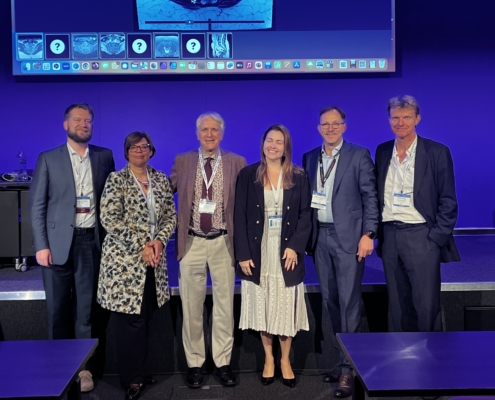 https://berlincaseviewer.de/wp-content/uploads/ISS-2023-faculty-landscape-scaled.jpeg
1616
2560
Eva Reidemeister
https://berlincaseviewer.de/wp-content/uploads/2020/05/bcv-logo-1.svg
Eva Reidemeister2023-11-03 22:06:362023-11-04 01:54:13MSK 2023 Joint Meeting With Interactive Course on axSpA
https://berlincaseviewer.de/wp-content/uploads/ISS-2023-faculty-landscape-scaled.jpeg
1616
2560
Eva Reidemeister
https://berlincaseviewer.de/wp-content/uploads/2020/05/bcv-logo-1.svg
Eva Reidemeister2023-11-03 22:06:362023-11-04 01:54:13MSK 2023 Joint Meeting With Interactive Course on axSpAThis post is also available in: German

