Detection of COVID-19 on CT Scans: BerlinCaseViewer as a Training Tool during the Current Coronavirus Pandemic
The novel coronavirus SARS-CoV-2 currently presents medical professionals in many countries with an unprecedented challenge. It can lead to pneumonia in a considerable proportion of cases with a serious clinical course or even death in some cases.
Many questions have arisen, of which not nearly all can be answered at the time. Some are:
- What is the exact length of the incubation period?
- What possibilities are there to detect the disease even in asymptomatic cases?
- How can COVID-19 differ from other pulmonary diseases (including other viral pneumonias)?
When fatigue, coughing, shortness of breath, sputum and fever occur, it could be a case of COVID-19. Yet, these symptoms do not exclude other illnesses, such as the common flu. But to relieve hospital staff in this time, it is of utmost importance to identify patients with COVID-19 early on.
Diagnosing COVID-19
A real-time reverse transcription-polymerase chain reaction (rRT-PCR) from throat and nose swabs is the standard method used for testing. But in early stages of the pulmonary illness, false negative results are possible. This is attributed to the fact that the sensitivity of the test lies – varying from study to study – between 60 and 97 percent.¹ ²
Medical professionals can draw additional inferences from imaging diagnostics. Whereas conventional X-rays tend to show changes in advanced stages, and even then merely unspecific ones, a CT scan of the thorax is considered to be of more relevance in diagnosing COVID-19.³
Combining Testing Methods
Just upon suspicion or with only mild symptoms occurring, the Fleischner Society recommends to refrain from using CT scans. However, with the patient’s condition deteriorating or with an attested infection of COVID-19, the renowned specialist association dedicated to the diagnosis and treatment of diseases of the chest, supports the CT scanning of the lung. For the doctors working in hospitals the scans both facilitate easier decision-making and justification for their measures taken.
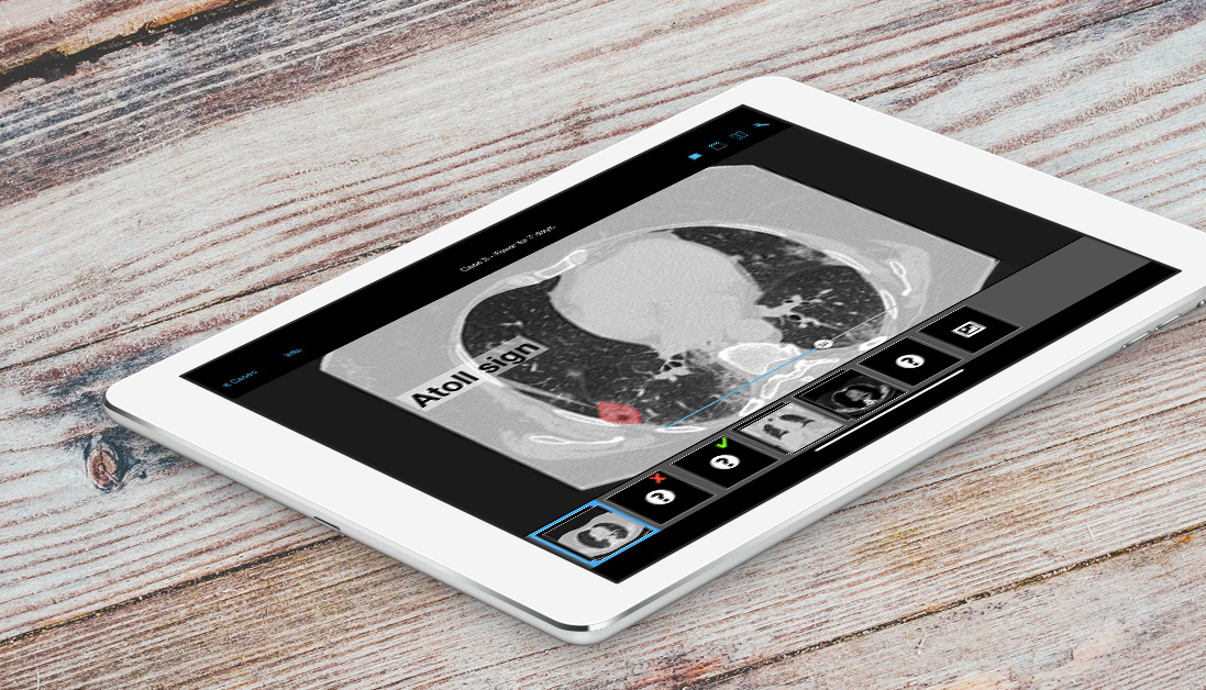
How to Detect COVID-19 on CT Scans?
CT scans are one way to detect COVID-19 in patients. That is, if the medical professionals from different fields – reaching from pulmonology and critical care medicine to radiology – are able to recognize the changes typically occurring in COVID-19 patients in the scans. At the same time they must be able to distinctly tell them apart from other inflammatory and non-inflammatory lung diseases.
Here are the phenomena to watch out for:
- Ground glass opacities
- Crazy paving pattern
- Air-space consolidation
Especially when the scans show peripheral occurence in the lung, or the signs appear in both lungs, as well as in the lower parts of the lung, then these could raise suspicion of COVID-19 in the appropriate clinical setting. However, even though the three indicators are relatively specific, they are still not pathognomonic. In any case, training is necessary to assess these saliences properly.
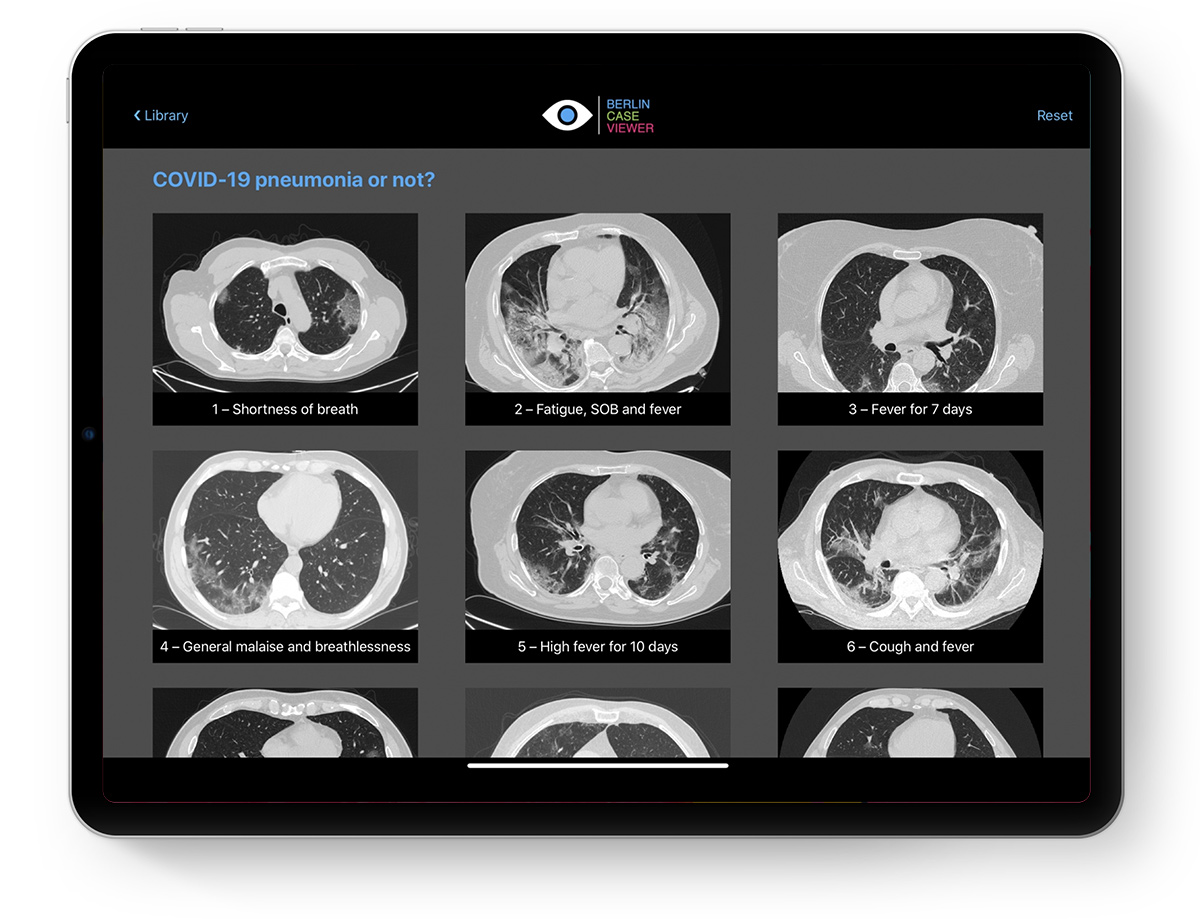
Coronavirus Pandemic: BerlinCaseViewer Focussing on COVID-19
This is where the BerlinCaseViewer (BCV) comes into play. The medical training application, usually focussing on rheumatic diseases, shifts its focus in times of the novel coronavirus on the new lung disease COVID-19.
Together with the radiologists Fabio Macori from Rome, Italy, and Yuranga Weerakkody from Perth, Australia, a quiz addressing the specific changes in the lungs of patients with COVID-19-related pneumonia was developed. The interactive e-learning tool also takes differential diagnoses into account. To make this possible, the radiologist Kay G. Hermann from the BerlinCaseViewer team brought together the experts for imaging of the lungs.
On the technical side of things, the BerlinCaseViewer app offered the ideal framework to showcase 19 interesting cases. The Italian image datasets were swiftly added to the BCV database and complemented with a handy quiz by using the integrated question generator.
About the Experts
As an acknowledged lung expert, the radiologist Fabio Macori served the Italian population with valuable imaging diagnostics. Yuranga Weerakkody is an internationally renowned radiologist from Australia with thoracic sub-specialty expertise and an enthusiasm for e-learning methods. He has been a voluntary contributor for radiopaedia.org for over a decade.

Fabio Macori

Yuranga Weerakkody
Our Quiz in Helping Radiologists and Clinicians during Coronavirus Crisis
With the new extension to the case collection of the app, doctors all over the world gain access to an e-learning tool to better understand the typical changes in the lungs with COVID-19. Get it now! The BerlinCaseViewer app for iPad can be downloaded for free in the AppStore.
This joint venture once more shows the importance of solidarity in difficult times. Modern technology enables us to realize such collaborations beyond national borders (#FightCovid) while working from home (#StayAtHome). With this in mind, we are hopeful to set bounds to SARS-CoV-2 across the globe.
References:
Download BerlinCaseViewer
Related posts:
 https://berlincaseviewer.de/wp-content/uploads/Rheuma-Day-2024-Header-WP.jpg
843
1500
Eva Reidemeister
https://berlincaseviewer.de/wp-content/uploads/2020/05/bcv-logo-1.svg
Eva Reidemeister2024-02-13 16:36:512024-02-13 16:36:51Clinical Insights: Rheuma Day 2024 — Focussing on MSK Radiology
https://berlincaseviewer.de/wp-content/uploads/Rheuma-Day-2024-Header-WP.jpg
843
1500
Eva Reidemeister
https://berlincaseviewer.de/wp-content/uploads/2020/05/bcv-logo-1.svg
Eva Reidemeister2024-02-13 16:36:512024-02-13 16:36:51Clinical Insights: Rheuma Day 2024 — Focussing on MSK Radiology https://berlincaseviewer.de/wp-content/uploads/ISS-2023-faculty-landscape-scaled.jpeg
1616
2560
Eva Reidemeister
https://berlincaseviewer.de/wp-content/uploads/2020/05/bcv-logo-1.svg
Eva Reidemeister2023-11-03 22:06:362023-11-04 01:54:13MSK 2023 Joint Meeting With Interactive Course on axSpA
https://berlincaseviewer.de/wp-content/uploads/ISS-2023-faculty-landscape-scaled.jpeg
1616
2560
Eva Reidemeister
https://berlincaseviewer.de/wp-content/uploads/2020/05/bcv-logo-1.svg
Eva Reidemeister2023-11-03 22:06:362023-11-04 01:54:13MSK 2023 Joint Meeting With Interactive Course on axSpA https://berlincaseviewer.de/wp-content/uploads/Version-3-LinkedIn-Social.jpeg
1080
1920
Eva Reidemeister
https://berlincaseviewer.de/wp-content/uploads/2020/05/bcv-logo-1.svg
Eva Reidemeister2023-10-04 22:02:582023-10-05 12:16:27Free Online Course on World Arthritis Day 2023: Understanding Imaging of Arthritis
https://berlincaseviewer.de/wp-content/uploads/Version-3-LinkedIn-Social.jpeg
1080
1920
Eva Reidemeister
https://berlincaseviewer.de/wp-content/uploads/2020/05/bcv-logo-1.svg
Eva Reidemeister2023-10-04 22:02:582023-10-05 12:16:27Free Online Course on World Arthritis Day 2023: Understanding Imaging of ArthritisThis post is also available in: German

